Etiketter

Summa sidvisningar

Sidor

Leta i den här bloggen

tisdag 8 oktober 2013

Fosfataasi entsyymifunktio on nykyään paremmin tiedossa

http://phys.org/news/2013-05-database-phosphate-substrate.html
It is now easier to pinpoint exactly what molecules a phosphatase – a type of protein that's essential for cells to react to their environment – acts upon in human cells, thanks to the free online database DEPOD, created by EMBL scientists. Published today in Science Signaling, the overview of interactions could even help explain unforeseen side-effects of drugs.
Although we know the tool's general purpose, it can sometimes be difficult to tell if a specific pair of precision tweezers belongs to a surgeon or a master jeweller. It is now easier to solve similar conundrums about a type of protein that allows cells to react to their environment, thanks to scientists at the European Molecular Biology Laboratory (EMBL). Published today in Science Signaling, their work offers a valuable resource for other researchers.
Whether in your eye being hit by light, in your blood fighting off disease, or elsewhere throughout your body, cells have to react to changes in their environment. But first, a cell must 'know' the environment has changed. One of the ways in which that information is transmitted within the cell is through tags called phosphate ions, which are added to or removed from specific molecules depending on the exact message that has to be conveyed. The tools the cell uses to remove phosphate ions are proteins called phosphatases. But it's not always obvious what molecules – or substrates – a particular phosphatase acts upon.
ESIMERKKI 
Ajattele inositolifosfaattien  ja fosfoinositidien varaan  rakennettua kehomme  signaalijärjestelmää kuin hienona kellon koneistona! Jos sellaiseen mättää  inositolijärjestelmälle turtaa  muuta fosfaattia- tarkka metbolinen ja signaloiva ajastin tukkeutuu
--
"One of the biggest challenges in phosphatase research is finding substrates, and this is what our work supports," says Maja Köhn from EMBL in Heidelberg, Germany, who led the study. "We've made it easier to create hypotheses about the relationships between phosphatases and their substrates."
Xun Li, a post-doctoral student shared by Köhn's lab and those of Matthias Wilmanns at EMBL in Hamburg, Germany and Janet Thornton at EMBL-European Bioinformatics Institute (EMBL-EBI) in Hinxton, UK, compiled the most complete picture to date of all the phosphatases in , and their substrates. The scientists also grouped phosphatases into families, based on their three-dimensional structure, which can influence what molecules a phosphatase can act upon.
This information allows researchers to easily identify a phosphatase's known substrates, and suggest new substrates based on how similar it is to other phosphatases. The web-like overview of interactions could even help explain unforeseen side-effects of drugs designed to interfere with phosphatases or with their phosphate-adding counterparts, kinases. To enable others to make such connections, Köhn and colleagues have created a free online database, DEPOD.
"When people have unexpected results, this could be a place to find explanations," says Thornton, head of EMBL-EBI. "DEPOD combines a wealth of information that can be explored in a variety of ways, to make it useful not just to phosphatase researchers but to the wider community."


Read more at: http://phys.org/news/2013-05-database-phosphate-substrate.html#jCp
Vielä näkökohta polyfosfaateista

http://www.ncbi.nlm.nih.gov/pmc/articles/PMC2084301/
En vielä käännä suomeksi näistä mitään.
Ajattelen asiaa dieetin kannalta: Dieetin on annettava  molekyylejä, joista keho voi saada esiin näitä fosfaatteja moniin tarkoituksiinsa.  Fytiini- muoto  edullinen.

 http://www.ncbi.nlm.nih.gov/pmc/articles/PMC1356573/

Polyfosfaattien muodostuskyvyn tärkeydestä ja niiden funktioista -pohdittavaksi

http://en.wikipedia.org/wiki/Polyphosphate
Tämä on Wikipediasta. Siitä ei ole suomalaista versiota:

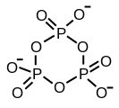
Triphosphates are salts or esters of polymeric oxyanions formed from tetrahedral PO4 (phosphate) structural units linked together by sharing oxygen atoms. When two corners are shared the polyphosphate may have a linear chain structure or a cyclic ring structure. In biology the polyphosphate esters AMP, ADP and ATP are involved in energy transfer. A variety of polyphosphates find application in mineral sequestration in municipal waters, generally being present at 1 to 5 pm.[1] GTP, CTP, and UTP are also nucleotides important in the protein synthesis, lipid synthesis and carbohydrate metabolism, respectively.

Structure & Formation

The structure of tripolyphosphoric acid illustrates the principles which define the structures of polyphosphates. It consists of three tetrahedral PO4 units linked together by sharing oxygen atoms. Structurally, the outer tetrahedra share one vertex with the central tetrahedron; the central tetrahedron shares two corners with the other tetrahedra. The corresponding phosphates are related to the acids by loss of the acidic protons. In the case of the cyclic trimer each tetrahedron shares two vertices with adjacent tetrahedra.
Sharing of three corners is possible as in the sheet-structure Phyllosilicates, but such structures occur only under extreme conditions. Three-corner sharing also occurs in phosphorus pentoxide, P4O10, which has a 3-dimensional structure.
Chemically, the polymerization reaction can be seen as a condensation reaction. The process begins with two phosphate units coming together.
2 PO43− + 2 H+ is in equilibrium with P2O74− + H2O
It is shown as an equilibrium reaction because it can go in the reverse direction, when it is known as an hydrolysis reaction because a water molecule is split (Lysed). The process may continue in steps; at each step another PO3 unit is added to the chain, as indicated by the part in brackets in the illustration of polyphosphoric acid. P4O10 can be seen as the end product of condensation reactions, where each tetrahedron shares three corners with the others. Conversely, a complex mix of polymers is produced when a small amount of water is added to phosphorus pentoxide.

Acid-base and complexation properties

Polyphosphates are weak bases. A lone pair of electrons on an oxygen atom can be donated to a hydrogen ion (proton) or a metal ion in a typical Lewis acid-Lewis base interaction. This has profound significance in biology. For instance, adenosine triphosphate (ATP)  is about 25% protonated in aqueous solution at pH 7.[2]
ATP4- + H+ is in equilibrium with ATPH3-, pKa \approx 6.6
Further protonation occurs at lower pH values.
ATP forms chelate complexes with metal ions. The stability constant for the equilibrium
ATP4- + Mg2+ is in equilibrium with MgATP2-, log β \approx 4
is particularly large.[3] The formation of the magnesium complex is a critical element in the process of ATP hydrolysis, as it weakens the link between the terminal phosphate group and the rest of the molecule.[2][4]

The "high energy" phosphate bond

The energy released in ATP hydrolysis,
ATP4- + H2O → ADP3- + Pi-
at ΔG \approx -36.8 kJ mol−1 is large by biological standards. Pi stands for inorganic phosphate, which is protonated at biological pH. However, it is not large by inorganic standards. The term "high energy" refers to the fact that it is high relative to the amount of energy released in the organic chemical reactions that can occur in living systems.

High-polymeric inorganic polyphosphates

High-polymeric inorganic polyphosphates were found in living organisms by L. Liberman in 1890. These compounds are linear polymers containing a few to several hundred residues of orthophosphate linked by energy-rich phosphoanhydride bonds.
Previously, it was considered either as “molecular fossil” or as only a phosphorus and energy source providing the survival of microorganisms under extreme conditions. These compounds are now known to also have regulatory roles, and to occur in representatives of all kingdoms of living organisms, participating in metabolic correction and control on both genetic and enzymatic levels. Polyphosphate is directly involved in the switching-over of the genetic program characteristic of the exponential growth stage of bacteria to the program of cell survival under stationary conditions, “a life in the slow line”. They participate in many regulatory mechanisms occurring in bacteria:
  • They participate in the induction of rpoS, an RNA-polymerase subunit which is responsible for the expression of a large group of genes involved in adjustments to the stationary growth phase and many stressful agents.
  • They are important for cell motility, biofilms formation and virulence.
  • Polyphosphates and exopolyphosphatases participate in the regulation of the levels of the stringent response factor, guanosine 5'-diphosphate 3'-diphosphate (ppGpp), a second messenger in bacterial cells.
  • Polyphosphates participate in the formation of channels across the living cell membranes. The above channels formed by polyphosphate and poly-b-hydroxybutyrate with Ca2+ are involved in the transport processes in a variety of organisms.
  • An important function of polyphosphate in microorganisms—prokaryotes and the lower eukaryotes—is to handle changing environmental conditions by providing phosphate and energy reserves. Polyphosphates are present in animal cells, and there are many data on its participation in the regulatory processes during development and cellular proliferation and differentiation—especially in bone tissues and brain.
In humans polyphosphates are shown to play a key role in blood coagulation. Produced and released by platelets[5]they activate Factor XII which is essential for blood clot formation. Furthermore platelets-derived polyphosphates activate blood coagulation factor XII (Hageman factor) that initiates fibrin formation and the generation of a proinflammatory mediator, bradykinin that contributes to leakage from the blood vessels and thrombosis.[6][7]

See also

Orgaanin polyfosfaatti solussa - mietittäväksi asiaksi

Chromosome replication and segregation govern the biogenesis and inheritance of inorganic polyphosphate granules
  1. Sean Crosson*,
+ Affiliations
  1. *Committee on Microbiology, University of Chicago, Chicago, IL 60637, USA
  2. †Department of Biochemistry and Molecular Biology, University of Chicago, Chicago, IL 60637 USA
  1. Fred Chang, Monitoring Editor
+ Affiliations
  1. Columbia University
  • Submitted April 5, 2013.
  • Revised August 15, 2013.
  • Accepted August 19, 2013.

Abstract

Prokaryotes and eukaryotes synthesize long chains of orthophosphate, known as polyphosphate (polyP), which form dense granules within the cell. PolyP regulates myriad cellular functions and is often localized to specific subcellular addresses through mechanisms that remain undefined. In this study, we present a molecular-level analysis of polyP subcellular localization in the model bacterium, Caulobacter crescentus. We demonstrate that biogenesis and localization of polyP is controlled as a function of the cell cycle, which ensures regular partitioning of granules between mother and daughter. The enzyme polyphosphate kinase 1 (Ppk1) is required for granule production, colocalizes with granules, and dynamically localizes to the sites of new granule synthesis in nascent daughter cells. Localization of Ppk1 within the cell requires an intact catalytic active site and a short, positively-charged tail at the C-terminus of the protein. The processes of chromosome replication and segregation govern both the number and position of Ppk1/polyP complexes within the cell. We propose a multi-step model whereby the chromosome establishes sites of polyP coalescence, which recruit Ppk1 to promote the in situ synthesis of large granules. These findings underscore the importance of both chromosome dynamics and discrete protein localization as organizing factors in bacterial cell biology.

Footnotes

This article has not yet been cited by other articles.

Hakusana Inositolipyrofosfaatit Inositolpyrophosphates

Katson 20  / 125:stä) uusimmasta tieteellisestä artikkelista.

Results: 1 to 20 of 125

1.
Kilari RS, Weaver JD, Shears SB, Safrany ST.
FEBS Lett. 2013 Sep 8. doi:pii: S0014-5793(13)00664-9. 10.1016/j.febslet.2013.08.035. [Epub ahead of print]
PMID:
24021644
[PubMed - as supplied by publisher]
2.
Erneux C, Elong Edimo W.
Biochem J. 2013 Aug 1;453(3):e3-4. doi: 10.1042/BJ20130785.
PMID:
23849059
[PubMed - indexed for MEDLINE]
3.
Ye C, Bandara WM, Greenberg ML.
J Biol Chem. 2013 Aug 23;288(34):24898-908. doi: 10.1074/jbc.M113.493353. Epub 2013 Jul 2.
PMID:
23824185
[PubMed - in process]
4.
Ghosh S, Shukla D, Suman K, Lakshmi BJ, Manorama R, Kumar S, Bhandari R.
Blood. 2013 Aug 22;122(8):1478-86. doi: 10.1182/blood-2013-01-481549. Epub 2013 Jun 19.
PMID:
23782934
[PubMed - in process]
5.
Wilson MS, Livermore TM, Saiardi A.
Biochem J. 2013 Jun 15;452(3):369-79. doi: 10.1042/BJ20130118. Review.
PMID:
23725456
[PubMed - indexed for MEDLINE]
6.
Worley J, Luo X, Capaldi AP.
Cell Rep. 2013 May 30;3(5):1476-82. doi: 10.1016/j.celrep.2013.03.043. Epub 2013 May 2.
PMID:
23643537
[PubMed - in process]
Free PMC Article
7.
Barker CJ, Berggren PO.
Pharmacol Rev. 2013 Feb 19;65(2):641-69. doi: 10.1124/pr.112.006775. Print 2013 Apr. Review.
PMID:
23429059
[PubMed - indexed for MEDLINE]
8.
Niger C, Luciotti MA, Buo AM, Hebert C, Ma V, Stains JP.
J Bone Miner Res. 2013 Jun;28(6):1468-77. doi: 10.1002/jbmr.1867.
PMID:
23322705
[PubMed - in process]
9.
Jadav RS, Chanduri MV, Sengupta S, Bhandari R.
J Biol Chem. 2013 Feb 1;288(5):3312-21. doi: 10.1074/jbc.M112.396556. Epub 2012 Dec 19.
PMID:
23255604
[PubMed - indexed for MEDLINE]
Free PMC Article
10.
Banfic H, Bedalov A, York JD, Visnjic D.
J Biol Chem. 2013 Jan 18;288(3):1717-25. doi: 10.1074/jbc.M112.412288. Epub 2012 Nov 24.
PMID:
23179856
[PubMed - indexed for MEDLINE]
11.
Shears SB, Weaver JD, Wang H.
Adv Biol Regul. 2013 Jan;53(1):19-27. doi: 10.1016/j.jbior.2012.10.002. Epub 2012 Oct 11. Review.
PMID:
23107997
[PubMed - indexed for MEDLINE]
12.
Wundenberg T, Mayr GW.
Biol Chem. 2012 Sep;393(9):979-98. doi: 10.1515/hsz-2012-0133. Review.
PMID:
22944697
[PubMed - indexed for MEDLINE]
13.
Barker CJ, Berggren PO.
Adv Biol Regul. 2012 Sep;52(3):361-8. doi: 10.1016/j.jbior.2012.05.002. Epub 2012 May 26. Review.
PMID:
22884029
[PubMed - indexed for MEDLINE]
14.
Saiardi A.
Adv Biol Regul. 2012 May;52(2):351-9. doi: 10.1016/j.jbior.2012.03.002. Epub 2012 Apr 5. Review.
PMID:
22781748
[PubMed - indexed for MEDLINE]
15.
Saiardi A.
Subcell Biochem. 2012;59:413-43. doi: 10.1007/978-94-007-3015-1_14.
PMID:
22374099
[PubMed - in process]
16.
Wang H, Falck JR, Hall TM, Shears SB.
Nat Chem Biol. 2011 Nov 27;8(1):111-6. doi: 10.1038/nchembio.733.
PMID:
22119861
[PubMed - indexed for MEDLINE]
17.
Szijgyarto Z, Garedew A, Azevedo C, Saiardi A.
Science. 2011 Nov 11;334(6057):802-5. doi: 10.1126/science.1211908.
PMID:
22076377
[PubMed - indexed for MEDLINE]
Free Article
18.
Loss O, Azevedo C, Szijgyarto Z, Bosch D, Saiardi A.
J Vis Exp. 2011 Sep 3;(55):e3027. doi: 10.3791/3027.
PMID:
21912370
[PubMed - indexed for MEDLINE]
Free PMC Article
19.
Chakraborty A, Kim S, Snyder SH.
Sci Signal. 2011 Aug 23;4(188):re1. doi: 10.1126/scisignal.2001958. Review.
PMID:
21878680
[PubMed - indexed for MEDLINE]
Free PMC Article
20.
Lonetti A, Szijgyarto Z, Bosch D, Loss O, Azevedo C, Saiardi A.
J Biol Chem. 2011 Sep 16;286(37):31966-74. doi: 10.1074/jbc.M111.266320. Epub 2011 Jul 20.
PMID:
21775424
[PubMed - indexed for MEDLINE]
Free PMC Article