Etiketter

Summa sidvisningar

Sidor

Leta i den här bloggen

måndag 28 oktober 2019

Fosfolipidit autofagosomin muodostuksessa ja fuusiossa


Journal of Molecular Biology
Volume 428, Issue 24, Part A, 4 December 2016, Pages 4819-4827
Review
Phospholipids in Autophagosome Formation and Fusion
ShuheiNakamura2TamotsuYoshimori2
Under a Creative Commons license

https://www.sciencedirect.com/science/article/pii/S0022283616304557

Highlights

Phosphoinositides (PI)  regulate the initiation and elongation of isolation membranes.
Lipids with small headgroups and, in particular, phosphatidylethanolamine (PE)  are likely to play important roles during autophagosome formation.
The generation and specific turnover of phosphoinositides (PI) on autophagosomal and lysosomal membranes regulate fusion.
The lipid composition of isolation membranes and autophagosomes is currently unknown.
 

Abstract

Autophagosomes are double-membrane organelles that are formed during a process referred to as macroautophagy. They serve to deliver cytoplasmic material into the lysosome for degradation. Autophagosomes are formed in a de novo manner and are the result of substantial membrane remodeling processes involving numerous protein–lipid interactions. While most studies focus on the proteins involved in autophagosome formation, it is obvious that lipids including phospholipids, sphingolipids, and sterols play an equally important role. Here, we summarize the current knowledge about the role of lipids, especially focusing on phospholipids and their interplay with the autophagic protein machinery during autophagosome formation and fusion.

lördag 26 oktober 2019

PI3K säätelyllinen alayksikkö silppuroituu BTB- Kelch perheen proteiinista KBTBD2

https://www.ncbi.nlm.nih.gov/gene/25948

Also known as
BKLHD1
Expression
Ubiquitous expression in gall bladder (RPKM 16.7), bone marrow (RPKM 14.7) and 25 other tissues See more
2016 Oct 18;113(42):E6418-E6426. Epub 2016 Oct 5.
Insulin resistance and diabetes caused by genetic or diet-induced KBTBD2 deficiency in mice.
We describe a metabolic disorder characterized by lipodystrophy, hepatic steatosis, insulin resistance, severe diabetes, and growth retardation observed in mice carrying N-ethyl-N-nitrosourea (ENU)-induced mutations. The disorder was ascribed to a mutation of kelch repeat and BTB (POZ) domain containing 2 (Kbtbd2) and was mimicked by a CRISPR/Cas9-targeted null allele of the same gene. Kbtbd2 encodes a BTB-Kelch family substrate recognition subunit of the Cullin-3-based E3 ubiquitin ligase. KBTBD2 targeted p85α, the regulatory subunit of the phosphoinositol-3-kinase (PI3K) heterodimer, causing p85α ubiquitination and proteasome-mediated degradation. In the absence of KBTBD2, p85α accumulated to 30-fold greater levels than in wild-type adipocytes, and excessive p110-free p85α blocked the binding of p85α-p110 heterodimers to IRS1, interrupting the insulin signal. Both transplantation of wild-type adipose tissue and homozygous germ line inactivation of the p85α-encoding gene Pik3r1 rescued diabetes and hepatic steatosis phenotypes of Kbtbd2-/- mice. Kbtbd2 was down-regulated in diet-induced obese insulin-resistant mice in a leptin-dependent manner. KBTBD2 is an essential regulator of the insulin-signaling pathway, modulating insulin sensitivity by limiting p85α abundance.

KEYWORDS:

Kbtbd2; diabetes; insulin resistance; p85α; ubiquitination

torsdag 3 oktober 2019

Aivojen alueen lipidistruktuurissa on havaittu AD taudissa varhain kardiolipiinin muutoksia synaptisissa mitokondrioissa.

https://www.ncbi.nlm.nih.gov/pubmed/25182746

Kardiolipiini eroaa  muista fosfolipidirakenteista olennaisesti ja sen toimintapaikka on tosiaankin  vain mitokodnria, jolla on erikoinen kalvorakenne, ulko kalvo ja sisäkalvo ja niitten välille jäänyt  enregiatarkoituksiin tärkeä  tila. Ulkokalvolla ja sisäkalvolla on  erilaiset fosfolipidikoostumat.
KARDIOLIPIINI  on tosi moduli joka on kondensoitu kolmesta  glyserolirungosta. R tarkoittaa rasvahappoa.
Kuva  Bildresultat för Cardiolipin

https://www.ncbi.nlm.nih.gov/pubmed/25627476
 https://link.springer.com/article/10.1007%2Fs10863-015-9607-y

  • Metabolisia mitokondrioita on tutkittu 2014, kardiolipiini, joka on mitokondrain sisäkalvossa.  Kardiolipiinirakenteeseen vaikuttaa ihmisen elintapa ja dieetti ainakin osaltaan,  herediteetti on tietysti  vahvin vaikuttaja. muta ihminen voi  elintavoillaan haitata kardiolipiinirakenteen  pätevyyttä. Ala on yksi linkki, josas on kuvattu mitokondrian ulompaa kaksoislipidikerrosta joka suojaa  sisäossaa, jonka ympärillä on poimuinen lipidikerros, ja siinä sisäkerroksessa on kardiolipiinin sijaintipaikka.  Siihen sisäkerrokseen asettu kellumaan  tämän "energialaitoksen" proteiiniketju, "hengitysketju" , joka  kuljttaa elektroneja ja ottaa vastaan happea, tekee   radikaalimuotoista  happea  ja muodostaa  vesimolekyyliä, metabolista  vettä H2O.  ATP-pakkauksia tekevä järjestelmä sijaitsee myös  siinä.  Tuottuvia protoneja  syötetään kalvovälitilaan ja sieltä ne virtaavat  alas ja  nergia otetaan fosfaattisidoksiin  proiteiinissa : ATP-pakkauksiin.  
  •  Alustana  tälle järjestelmälle täytyy olla  normaalia kardiolipiiniä.

2014 Oct;46(5):447-57. doi: 10.1007/s10863-014-9555-y. Epub 2014 Jun 21.
Impact of high dietary lipid intake and related metabolic disorders on the abundance and acyl composition of the unique mitochondrial phospholipid, cardiolipin.
UMR 866, Dynamique Musculaire et Métabolisme, Centre INRA de Montpellier, 34060, Montpellier,Abstract
Excessive dietary lipid intake, coupled with lack of exercise, are the major causes of the development and progression of metabolic syndrome features e. g. obesity, hepatic steatosis, insulin resistance, type 2 diabetes and cardiovascular diseases. These metabolic diseases are associated with both structural and functional alterations of mitochondria.  
Cardiolipin (CL) is a unique phospholipid that is almost exclusively localized in the mitochondrial inner membrane.

 Cardiolipin is at the heart of mitochondrial metabolism playing a key role in several processes of mitochondrial bioenergetics as well as in mitochondrial membrane stability and dynamics, and in many of the mitochondrial-dependent steps of apoptosis. Indeed, alterations to CL content and acyl chain profile have been associated with mitochondrial dysfunction in multiple tissues in Barth syndrome and in many other physio-pathological conditions. After a brief overview of the biological roles of CL, we highlight the consequences of lipid overload-related nutritional manipulations as well as related metabolic disorders on both CL content and its fatty acid composition in the major metabolic tissues, the heart, muscle and liver. The goal of this review is to fill a void in the CL literature concerning the effects of CL abundance and form that arise following high lipid supplementation and the related metabolic disorders.
[Indexed for MEDLINE]
https://qph.fs.quoracdn.net/main-qimg-17d708fb1ead71db48b33e79ec6a05bf-c 


  •  Viime vuodelta löytyy selvitystä kardiolipidin molekulaarisesta  koostumuskesta. 
https://www.ncbi.nlm.nih.gov/pubmed/29618609
2018 Apr 17;115(16):4158-4163. doi: 10.1073/pnas.1719407115. Epub 2018 Apr 4.

Molecular structural diversity of mitochondrial cardiolipins.Oemer G1, Lackner K1, Muigg K1, Krumschnabel G2, Watschinger K3, Sailer S3, Lindner H4, Gnaiger E2, Wortmann SB5,6, Werner ER3, Zschocke J1, Keller MA7.Abstract

cardiolipin; lipids; mass spectrometry; mathematical modeling; mitochondria
PMID:
29618609
PMCID:
PMC5910844
DOI:
10.1073/pnas.1719407115
[Indexed for MEDLINE]
Free PMC Article
  • ENTÄ SITtEN AIVOJEN mitokondrioitten kardiolipiinin piirteet?
     

    2015;43(4):1375-92. doi: 10.3233/JAD-141002.

    Cardiolipin profile changes are associated to the early synaptic mitochondrial dysfunction in Alzheimer's disease.

    l. Abstract
    Brain mitochondria are fundamental to maintaining healthy functional brains, and their dysfunction is involved in age-related neurodegenerative disorders such as Alzheimer's disease (AD). In this study, we conducted a research on how both non-synaptic and synaptic mitochondrial functions are compromised at an early stage of AD-like pathologies and their correlation with putative changes on membranes lipid profile, using 3 month-old nontransgenic and 3xTg-AD mice, a murine model of experimental AD. Bioenergetic dysfunction in 3xTg-AD brains is evidenced by a decrease of brain ATP levels resulting, essentially, from synaptic mitochondria functionality disruption as indicated by declined respiratory control ratio associated with a 50% decreased complex I activity. Lipidomics studies revealed that synaptic bioenergetic deficit of 3xTg-AD brains is accompanied by alterations in the phospholipid composition of synaptic mitochondrial membranes, detected either in phospholipid class distribution or in the phospholipids molecular profile. Globally, diacyl- and lyso-phosphatidylcholine lipids increase while ethanolamine plasmalogens and cardiolipins content drops in relation to nontransgenic background
     However, the main lipidomic mark of 3xTg-AD brains is that cardiolipin cluster-organized profile is lost in synaptic mitochondria due to a decline of the most representative molecular species.
    In contrast to synaptic mitochondria, results support the idea that non-synaptic mitochondria function is preserved at the age of 3 months. Although the genetically construed 3xTg-AD mouse model does not represent the most prevalent form of AD in humans, the present study provides insights into the earliest biochemical events in AD brain, connecting specific lipidomic changes with synaptic bioenergetic deficit that may contribute to the progressive synapses loss and the neurodegenerative process that characterizes AD. KEYWORDS:
    Alzheimer's disease; brain bioenergetics; cardiolipin; mitochondrial lipidomics.

Jos PICALM toimii hyvin, mikä on PIP2:n rasvahappokoosumus hermokudoksessa? Steariinihaposta

PICALM  kiinnittyy PIP2:n kalvossa kohdallaan jossa on lysiinejä K kolme  ja yksi histidiini H. Lysiini on essentielli aminohappo ja histidiini on joissain tapauksissa essentielli aminohappo.
PIP2 rakenne on modulimainen ja kaikissa PIP2 muodoissa on  inositolirengas, ja siinä  fosfaatteja sekä fosfatidyyli, jossa on  kaksi rasvahappoa. Tämä fosfatidyylikohta on kuin se varsi, jolla moduli on petautunut palsamkalvolipidien joukkoon.
Näillä  rasvahapoilla on myös geneettinen preferenssinsä.   Glyserolirakenne joka on  pohjilla  näissä  moduleissa, ottaa sn1 asemaan mieluyiten steariinihappoa, jota keho tuottaa etikkahaposta käsin palmitiinihapon kautta. Sn2 asemaan  valikoituu mieluiten essentielli aminohapposyntyinen arakidonihapp . Sitä keho tekee  ravinnon  linolihapost käsin.  ja sitä onkin  siten kehon joka solussa normaalisti.
Mikä preferenssi on PIP2 modulin kokoontumsiessa  neurologisessa kudoksessa, onko   se tämä  sama yleinen?   Tietysti näitä tutkimuksia on tehtykin paljon ja niitä vain tulee löytää.  Itselläkin pitäisi josain vihossa olla  luettelo   tästä eri fosfolipidien  tavasta valikoida   rakenteensa  rasvahapot.

https://en.wikipedia.org/wiki/Phosphatidylinositol
The specific fatty acids of PtdIns, and their conformation, employed in the sensory neurons has not been elucidated.  (Aug 2018).
 ----

Steariinihapon synteesi tapahtuu etikkahaposta (C2:0)   käsin  C18:0 kokoon.

 (Steariinihaposta  4 artikkelia):

https://www.ncbi.nlm.nih.gov/pubmed/28757255 
https://www.ncbi.nlm.nih.gov/pubmed/21732666 

2004 Apr 20;5:15. https://www.ncbi.nlm.nih.gov/pubmed/15099403 Regulation of stearoyl-CoA desaturase-1 after central and peripheral nerve lesions.Breuer S1, Pech K, Buss A, Spitzer C, Ozols J, Hol EM, Heussen N, Noth J, Schwaiger FW, Schmitt AB. Department of Neurology, University Medical School, Pauwelsstr 30, D-52074 Aachen, Germany.
Abstract BACKGROUND:
Interruption of mature axons activates a cascade of events in neuronal cell bodies which leads to various outcomes from functional regeneration in the PNS to the failure of any significant regeneration in the CNS. One factor which seems to play an important role in the molecular programs after axotomy is the stearoyl Coenzyme A-desaturase-1 (SCD-1). This enzyme is needed for the conversion of stearate (C18:0)  into oleate (C18:1). Beside its role in membrane synthesis, oleate could act as a neurotrophic factor, involved in signal transduction pathways via activation of protein kinases C. RESULTS: In situ hybridization and immunohistochemistry demonstrated a strong up-regulation of SCD at mRNA and protein level in regenerating neurons of the rat facial nucleus whereas non-regenerating Clarke's and Red nucleus neurons did not show an induction of this gene. CONCLUSION: This differential expression points to a functionally significant role for the SCD-1 in the process of regeneration. PMID: 15099403 PMCID: PMC411035 DOI: 10.1186/1471-2202-5-15 [Indexed for MEDLINE]

 https://www.ncbi.nlm.nih.gov/pubmed/7463084
Myeliinin gangliosidit  kananpojilla ja  kyyhkysillä  on tutkittu ja  rasvahappokoostumuksia verrattu  imettäväisten  gangliosideihin.  Steariinihappo oli yleinen  rasvahapporyhmässä,  10 prosenttia C20.-sfingosiinista.

Fatty acids of this ganglioside included both the hydroxy- and unsubstituted types, and long-chain bases were almost entirely C18. Ganglioside GM1 split into two closely migrating bands on TLC, the slower of which resembled mammalian GM1 in having stearate as the main fatty acid with a measurable amount (10%) of C20-sphingosine;


  • Entä jos puuttuu SCD1 aktiivisuus, eikä  stearaatti voi muuttua öljyhapoksi hermokudoksessa?
  •  Leptiinin ( fysiologisesti ilmeisesti )  tulisi aktivoida SCD1  suorittamaan konversio. 

 https://www.ncbi.nlm.nih.gov/pubmed/28768997/

  • Leptiiniresistenssi?

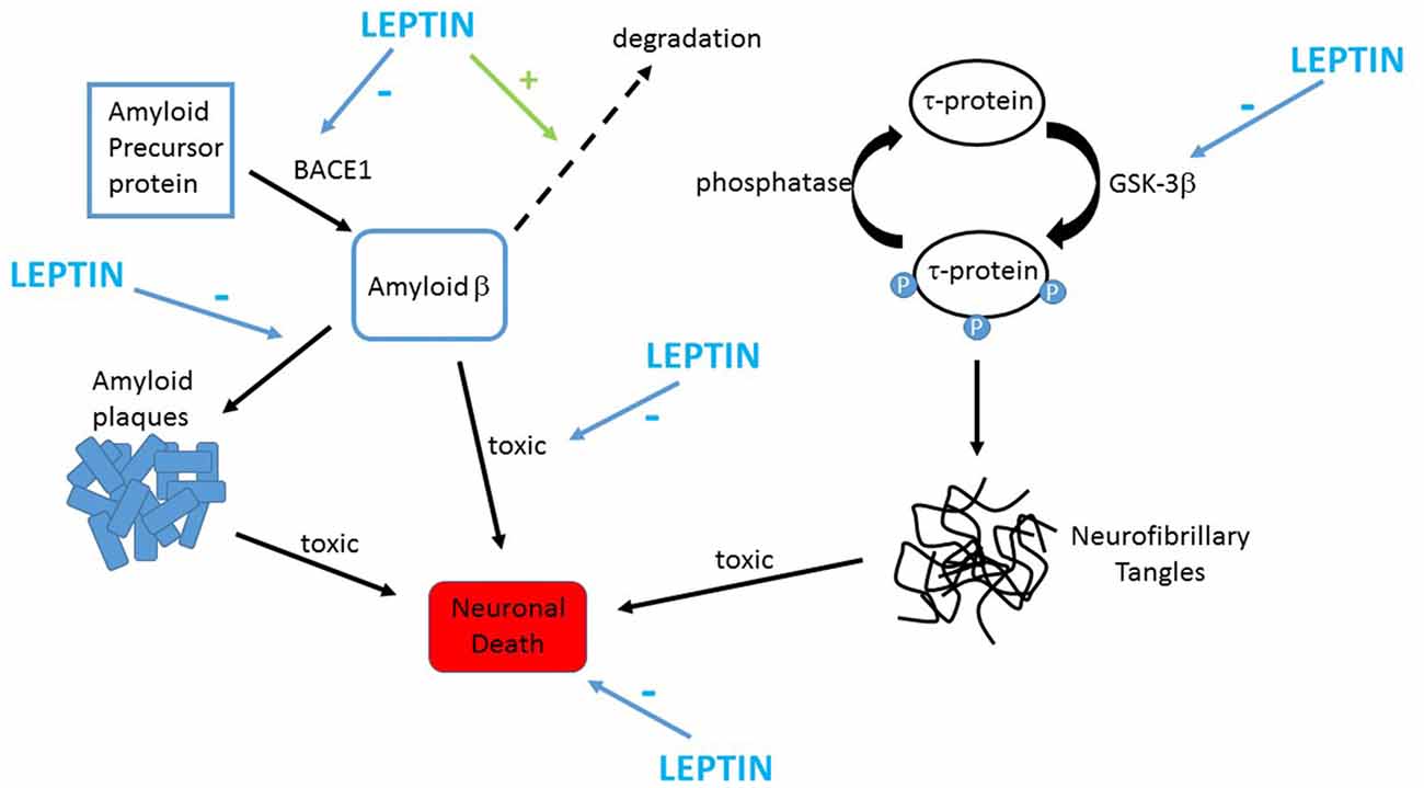
Leptiini ja neurodegeneraatio?
 https://www.ncbi.nlm.nih.gov/pubmed/?term=leptin%2C+neurodegeneration

https://www.sciencedirect.com/science/article/pii/S0028390817306226?via%3Dihub

 Tässä artikkeli joka kannattaa  suomentaakin.

 https://www.frontiersin.org/files/Articles/413613/fncel-12-00340-HTML/image_m/fncel-12-00340-g002.jpg

 https://www.frontiersin.org/files/Articles/413613/fncel-12-00340-HTML/image_m/fncel-12-00340-g002.jpg


...steariinihappopointsu..  Jatkuu.. 3.10.2019

Huom. ajattele steariinikynttilöitä. Steariinihappo on kovaa rasvaa. Leptiini- jos se toimii,   stimuloi sen muuttumista ( desaturoitumista)  MUFA hapoksi, joka on  öljyhappo.

LEPTIINI  vaikuttaa olevan  aika olennaisessa kohdasas toimiva peptidi "Obesity factor"- jos sen toiminta  vikuuntuu, aiheutuu ylipainoa ja painonvakauden menetys,  diabeettisuutat (DM2) ym.

Se vaikuttaa myös että Abeeta degradoituu, joten  sen vikatoiminta  on tekijä myös  Ad-taudissa.

Otan sivuhypyn:

Leptiinireseptori ja leptiini.


PICALM, fosfatidyyli-inositolia sitova klatriinia koostava proteiini

https://www.youtube.com/watch?v=QzGrvCHgQO0
https://www.ncbi.nlm.nih.gov/gene/8301 
Official Symbol
PICALMprovided by HGNC
Official Full Name
phosphatidylinositol binding clathrin assembly proteinprovided by HGNC
Also known as
LAP; CALM; CLTH
Preferred Names
phosphatidylinositol-binding clathrin assembly protein
Names
clathrin assembly lymphoid myeloid leukemia protein
Summary
This gene encodes a clathrin assembly protein, which recruits clathrin and adaptor protein complex 2 (AP2) to cell membranes at sites of coated-pit formation and clathrin-vesicle assembly. The protein may be required to determine the amount of membrane to be recycled, possibly by regulating the size of the clathrin cage. The protein is involved in AP2-dependent clathrin-mediated endocytosis at the neuromuscular junction. A chromosomal translocation t(10;11)(p13;q14) leading to the fusion of this gene and the MLLT10 gene is found in acute lymphoblastic leukemia, acute myeloid leukemia and malignant lymphomas. The polymorphisms of this gene are associated with the risk of Alzheimer disease. Multiple alternatively spliced transcript variants encoding different isoforms have been found for this gene. [provided by RefSeq, May 2011]
Expression
Ubiquitous expression in fat (RPKM 64.4), appendix (RPKM 52.9) and 25 other tissues See more
Orthologsmouse all


Bibliography


 https://www.ncbi.nlm.nih.gov/pubmed/30472946

 We found statistically significant association between the AD risk variant PICALM GG and increase in the P3 latency in subjects over 50 years old. The age-dependent increase in the P3 latency was more pronounced in the PICALM GG carriers than in the carriers of the PICALM AA and PICALM AG genotypes.

 https://www.ncbi.nlm.nih.gov/protein/NP_001008660.1


                /organism="Homo sapiens"
                     /db_xref="taxon:9606"
                     /chromosome="11"
                     /map="11q14.2"
     Protein         1..610
                     /product="phosphatidylinositol-binding clathrin assembly
                     protein isoform 2"
                     /note="clathrin assembly lymphoid myeloid leukemia
                     protein"
                     /calculated_mol_wt=66262
     Region          24..282
                     /region_name="ANTH"
                     /note="ANTH domain; pfam07651"
                     /db_xref="CDD:311541"
     Site            order(28,38,40..41)
                     /site_type="other"
                     /note="PtdIns(4,5)P2-binding site"
                     /db_xref="CDD:239622"
     Region          <268 ..="">366
                     /region_name="COG4223"
                     /note="Uncharacterized conserved protein [Function
                     unknown]"
                     /db_xref="CDD:226676"
     CDS             1..610
                     /gene="PICALM"
                     /gene_synonym="CALM; CLTH; LAP"
                     /coded_by="NM_001008660.3:305..2137"
                     /note="isoform 2 is encoded by transcript variant 2"
                     /db_xref="CCDS:CCDS31653.1"
                     /db_xref="GeneID:8301"
                     /db_xref="HGNC:HGNC:15514"
                     /db_xref="MIM:603025"
ORIGIN      
        1 msgqsltdri taaqhsvtgs avsktvckat theimgpkkk hldyliqctn emnvnipqla
       61 dslferttns swvvvfksli tthhlmvygn erfiqylasr ntlfnlsnfl dksglqgydm
      121 stfirrysry lnekavsyrq vafdftkvkr gadgvmrtmn tekllktvpi iqnqmdalld
      181 fnvnsneltn gvinaafmll fkdairlfaa ynegiinlle kyfdmkknqc kegldiykkf
      241 ltrmtrisef lkvaeqvgid rgdipdlsqa psslldaleq hlaslegkki kdstaasrat
      301 tlsnavssla stglsltkvd erekqaalee eqarlkalke qrlkelakkp htslttaasp
      361 vstsaggimt apaidifstp sssnstsklp ndlldlqqpt fhpsvhpmst asqvastwgg
      421 ftpspvaqph psaglnvdfe svfgnkstnv ivdsggfdel ggllkptvas qnqnlpvakl
      481 ppsklvsddl dsslanlvgn lgigngttkn dvnwsqpgek kltggsnwqp kvapttawna
      541 atmngmhfpq yappvmaypa ttptgmigyg ippqmgsvpv mtqptliysq pvmrppnpfg
      601 pvsgaqiqfm
//

(PIP2 koostumuksen  merkitys: PIP2:n  rasvahappokoostumus  on riippuvainen ravinnosta.
Fosftidyyli-inositolit valitsevat ensisijassa fosfatidyyliin rasvahappoja steariinihappo sn1 asemaan ja arakidonihappo sn2 asemaan.   Katson  tätä  PIP2;n  "kiinnitysvartta", pitkien  rasvahappojen kohtaa tarkemmin, varsinkin  steariinihaposta seuraavassa muistiinpanoa)