Etiketter

Summa sidvisningar

Sidor

Leta i den här bloggen

söndag 1 maj 2016

Insuliinireistenssisignaalitie

http://www.ncbi.nlm.nih.gov/pubmed/27102788


J Nutr Health Aging. 2016;20(5):509-13. doi: 10.1007/s12603-015-0601-1.

A High-sugar High-fat Diet Induced Metabolic Syndrome Shows some Symptoms of Alzheimer's Disease in Rats.

Abstract

OBJECTIVES:

Cases of sporadic Alzheimer's disease (SAD) are the predominant form of the age-related dementia. New evidence suggests that metabolic syndrome (MS), a metabolic disorder, is an initiating factor of some SAD cases. A high-sugar high-fat diet could cause MS, we aimed to investigate whether it could directly lead to SAD.

MEASUREMENTS:

The characteristic molecules of AD (hippocampus Aβ and Tau) were tested by using ELISA and western blotting to confirm the happening hallmarks of AD in brain. MS and inflammation related biochemical indicators were measured using immunological method. Proteins associated with the insulin resistance signal pathway (JNK, PI-3K, AKT, GSK-3β, GLUT3) were evaluated using western blotting method. The levels of reactive oxygen species (ROS) were measured by immunofluorescence method.

RESULTS:

Expressions of hippocampus Aβ, phosphorylation-Tau (p-Tau), inflammatory factors and p-JNK, Gsk-3βwere higher in the model rats than those in the control rats and expressions of p-PI3K, p-AKT and GLUT3 were reversed.

CONCLUSIONS:

The MS model animals, which can induce the characteristics symptoms of AD, and therefore it may be preliminarily considered that the AD pertains to the MS-related diseases.
PMID:
27102788
[PubMed - in process]
Tässä insuliiniresistenssissä    näen mahdollisen kohdan, jossa E-vitamiinin aineenvaihdunnallinen kit omaa  paikkansa normalisoimassa insuliiniresitenssiä. - vaikuttaa siltä ettäsiiehn kuuluu tasapainoisesti kolesterolilinjan molekyylit - siis kehon  isoprenoidist omat rakenteet   ja  ainakin A-vitamiini ja E vitamiini ravintoperäisinä, koska ne tekevät interferenssiä  fosfaatin  käyttöön ( ja on- off järejstelmään)
 
 Sen takia  verensokerin normalisoimisen ohella, dieetin tasaapinottamisen ohella ( fytiinin osa korostetuksi)  verernlipidien tasaapinottaminen on olennaista.  mutta ratkaisu ei ole vain jokin alentava  lääke ja endogeenit synteesitiet laajalti  blokeeraava mekanismi ja  epäselektiivisti kehosta rasvat poistava preparaatti. Pitäisi löytää sepsifisempia  metodeja ja tietysti  tarkentaa ravinto ja liikkuminen tarkemmin kontrolloidusti- esim kehon rasvojen sijainti ja  vaste liikuntaan-ja mistä  liikarasvoittuminen kiikastaa- koska  on paljon niitä,jotka "eivät mielestään  syö mitään# ja  "sittenkin lihovat" ja niitäm jotka noudttavat ohjeen toisensa jälkeen ja  eivät saa  veriarvoja normalisoitumaan tai  kehoaan  vaikuttamaan rterveeltä. Mutta onhan tähänsiten  tuleville sukupolville sadat vuodet  jatkoteesien  tekemiseen  aikaa. kehonpaino sinänsä ei heijasta liikarasvoittumista. Liikarasvainenvoinolla sitenkin, että ihan kelluu vedessä ja paino on poissa lihaksista ja luusta.  Impedanssimenetelmällä voi selvittää tämän  hämäävän "liikalihavuuden"  AD potilailla sataa hyväkuntoisuus olla sokeria ja rasvaa ja proteiinit puuttuvat, muta asia ei näy.

tokoferoliin assosioituva proteiini TAP

the human tocopherol-associated protein 1 (hTAP1/SEC14L2)
 https://archive.org/details/pubmed-PMC4077815

Viallinen alfaTTP-PIP-kompleksi ja perinnällinen E-vitamiinivaje

Science. 2013 May 31;340(6136):1106-10. doi: 10.1126/science.1233508. Epub 2013 Apr 18.

Impaired α-TTP-PIPs interaction underlies familial vitamin E deficiency.

Abstract

α-Tocopherol (vitamin E) transfer protein (α-TTP) regulates the secretion of α-tocopherol from liver cells. Missense mutations of some arginine residues at the surface of α-TTP cause severe vitamin E deficiency in humans, but the role of these residues is unclear. Here, we found that wild-type α-TTP bound phosphatidylinositol phosphates (PIPs), whereas the arginine mutants did not. In addition, PIPs in the target membrane promoted the intermembrane transfer of α-tocopherol by α-TTP. The crystal structure of the α-TTP-PIPs complex revealed that the disease-related arginine residues interacted with phosphate groups of the PIPs and that the PIPs binding caused the lid of the α-tocopherol-binding pocket to open. Thus, PIPs have a role in promoting the release of a ligand from a lipid-transfer protein.

Comment in

PMID:
23599266
[PubMed - indexed for MEDLINE]
Free full text

E-vitamiinia käsitelevä proteiini TAP

alpha-tocopherol transfer protein [Homo sapiens]

NCBI Reference Sequence: NP_000361.1
LOCUS       NP_000361                278 aa            linear   PRI 15-MAR-2015
DEFINITION  alpha-tocopherol transfer protein [Homo sapiens].
ACCESSION   NP_000361 XP_001128514
VERSION     NP_000361.1  GI:4507723
DBSOURCE    REFSEQ: accession NM_000370.3
KEYWORDS    RefSeq.
SOURCE      Homo sapiens (human)
  ORGANISM  Homo sapiens
            Eukaryota; Metazoa; Chordata; Craniata; Vertebrata; Euteleostomi;
            Mammalia; Eutheria; Euarchontoglires; Primates; Haplorrhini;
            Catarrhini; Hominidae; Homo.
REFERENCE   1  (residues 1 to 278)
  AUTHORS   Kono N, Ohto U, Hiramatsu T, Urabe M, Uchida Y, Satow Y and Arai H.
  TITLE     Impaired alpha-TTP-PIPs interaction underlies familial vitamin E
            deficiency
  JOURNAL   Science 340 (6136), 1106-1110 (2013)
   PUBMED   23599266
  REMARK    GeneRIF: The crystal structure of the
            alpha-TTP-phosphatidylinositol phosphates (PIPs) complex revealed
            that the familial vitamin E deficiency-related arginine residues
            interacted with phosphate groups of the PIPs and that the PIPs
            binding caused the lid of the alpha-tocopherol-binding pocket to
            open.
REFERENCE   2  (residues 1 to 278)
  AUTHORS   Etzl RP, Vrekoussis T, Kuhn C, Schulze S, Poschl JM, Makrigiannakis
            A, Jeschke U and Rotzoll DE.
  TITLE     Oxidative stress stimulates alpha-tocopherol transfer protein in
            human trophoblast tumor cells BeWo
  JOURNAL   J Perinat Med 40 (4), 373-378 (2012)
   PUBMED   22752767
  REMARK    GeneRIF: study demonstrated that alpha -TTP can be upregulated in
            case of oxidative stress in BeWo trophoblast cells; speculate that
            possibly, alpha -TTP is notonly involved in normal pregnancy, but
            also in cases of pregnancy disorders with intense oxidative stress
            Publication Status: Online-Only
REFERENCE   3  (residues 1 to 278)
  AUTHORS   Zhang WX, Thakur V, Lomize A, Pogozheva I, Panagabko C, Cecchini M,
            Baptist M, Morley S, Manor D and Atkinson J.
  TITLE     The contribution of surface residues to membrane binding and ligand
            transfer by the alpha-tocopherol transfer protein (alpha-TTP)
  JOURNAL   J. Mol. Biol. 405 (4), 972-988 (2011)
   PUBMED   21110980
  REMARK    GeneRIF: Substitution of residues in helices A8 (F165A and F169A)
            and A10 (I202A, V206A and M209A) decreased the rate of
            intermembrane ligand transfer as well as protein adsorption to
            phospholipid bilayers.
REFERENCE   4  (residues 1 to 278)
  AUTHORS   Booij JC, Bakker A, Kulumbetova J, Moutaoukil Y, Smeets B, Verheij
            J, Kroes HY, Klaver CC, van Schooneveld M, Bergen AA and Florijn
            RJ.
  TITLE     Simultaneous mutation detection in 90 retinal disease genes in
            multiple patients using a custom-designed 300-kb retinal
            resequencing chip
  JOURNAL   Ophthalmology 118 (1), 160-167 (2011)
   PUBMED   20801516
  REMARK    GeneRIF: Observational study of genetic testing. (HuGE Navigator)
REFERENCE   5  (residues 1 to 278)
  AUTHORS   Morley S, Thakur V, Danielpour D, Parker R, Arai H, Atkinson J,
            Barnholtz-Sloan J, Klein E and Manor D.
  TITLE     Tocopherol transfer protein sensitizes prostate cancer cells to
            vitamin E
  JOURNAL   J. Biol. Chem. 285 (46), 35578-35589 (2010)
   PUBMED   20826775
  REMARK    GeneRIF: Data show that reduction ('knockdown') of tocopherol
            transfer protein (TTP) expression resulted in resistance to the
            vitamin E.
REFERENCE   6  (residues 1 to 278)
  AUTHORS   Gotoda T, Arita M, Arai H, Inoue K, Yokota T, Fukuo Y, Yazaki Y and
            Yamada N.
  TITLE     Adult-onset spinocerebellar dysfunction caused by a mutation in the
            gene for the alpha-tocopherol-transfer protein
  JOURNAL   N. Engl. J. Med. 333 (20), 1313-1318 (1995)
   PUBMED   7566022
REFERENCE   7  (residues 1 to 278)
  AUTHORS   Arita M, Sato Y, Miyata A, Tanabe T, Takahashi E, Kayden HJ, Arai H
            and Inoue K.
  TITLE     Human alpha-tocopherol transfer protein: cDNA cloning, expression
            and chromosomal localization
  JOURNAL   Biochem. J. 306 (PT 2), 437-443 (1995)
   PUBMED   7887897
REFERENCE   8  (residues 1 to 278)
  AUTHORS   Ouahchi K, Arita M, Kayden H, Hentati F, Ben Hamida M, Sokol R,
            Arai H, Inoue K, Mandel JL and Koenig M.
  TITLE     Ataxia with isolated vitamin E deficiency is caused by mutations in
            the alpha-tocopherol transfer protein
  JOURNAL   Nat. Genet. 9 (2), 141-145 (1995)
   PUBMED   7719340
REFERENCE   9  (residues 1 to 278)
  AUTHORS   Ben Hamida C, Doerflinger N, Belal S, Linder C, Reutenauer L, Dib
            C, Gyapay G, Vignal A, Le Paslier D and Cohen D.
  CONSRTM   et al
  TITLE     Localization of Friedreich ataxia phenotype with selective vitamin
            E deficiency to chromosome 8q by homozygosity mapping
  JOURNAL   Nat. Genet. 5 (2), 195-200 (1993)
   PUBMED   8252047
REFERENCE   10 (residues 1 to 278)
  AUTHORS   Schuelke,M.
  TITLE     Ataxia with Vitamin E Deficiency
  JOURNAL   (in) Pagon RA, Adam MP, Ardinger HH, Bird TD, Dolan CR, Fong CT,
            Smith RJH and Stephens K (Eds.);
            GENEREVIEWS(R);
            (1993)
   PUBMED   20301419
COMMENT     REVIEWED REFSEQ: This record has been curated by NCBI staff. The
            reference sequence was derived from BC058000.1, BC041784.1 and
            AC120042.3.
            This sequence is a reference standard in the RefSeqGene project.
            On Sep 20, 2006 this sequence version replaced gi:113420428.
            
            Summary: This gene encodes a soluble protein that binds
            alpha-tocopherol, a form of vitamin E, with high selectivity and
            affinity. This protein plays an important role in regulating
            vitamin E levels in the body by transporting vitamin E between
            membrane vesicles and facilitating the secretion of vitamin E from
            hepatocytes to circulating lipoproteins. Mutations in this gene
            cause hereditary vitamin E deficiency (ataxia with vitamin E
            deficiency, AVED) and retinitis pigmentosa. [provided by RefSeq,
            Nov 2009].
            
            Sequence Note: This RefSeq record was created from transcript and
            genomic sequence data to make the sequence consistent with the
            reference genome assembly. The genomic coordinates used for the
            transcript record were based on transcript alignments.
            
            Publication Note:  This RefSeq record includes a subset of the
            publications that are available for this gene. Please see the Gene
            record to access additional publications.
            
            ##Evidence-Data-START##
            Transcript exon combination :: BC058000.1, BC041784.1 [ECO:0000332]
            RNAseq introns              :: single sample supports all introns
                                           SAMEA1968540, SAMEA1970526
                                           [ECO:0000348]
            ##Evidence-Data-END##
FEATURES             Location/Qualifiers
     source          1..278
                     /organism="Homo sapiens"
                     /db_xref="taxon:9606"
                     /chromosome="8"
                     /map="8q12.3"
     Protein         1..278
                     /product="alpha-tocopherol transfer protein"
                     /note="tocopherol (alpha) transfer protein (ataxia
                     (Friedreich-like) with vitamin E deficiency); alpha-TTP"
                     /calculated_mol_wt=31619
     Region          <40 ..73="" cdd="" cddsrv.cgi="" db_xref="CDD:<a href=" http:="" note="CRAL/TRIO, N-terminal domain; smart01100" region_name="CRAL_TRIO_N" tructure="" uid="215024" www.ncbi.nlm.nih.gov="">215024
" Region 95..248 /region_name="SEC14" /note="Sec14p-like lipid-binding domain. Found in secretory proteins, such as S. cerevisiae phosphatidylinositol transfer protein (Sec14p), and in lipid regulated proteins such as RhoGAPs, RhoGEFs and neurofibromin (NF1). SEC14 domain of Dbl is known to...; cd00170" /db_xref="CDD:238099" Site order(113,115,117,143,158,160,176,180,184,188,191,194,196, 203,210,222) /site_type="other" /note="phospholipid binding pocket [chemical binding]" /db_xref="CDD:238099" Site order(189,221) /site_type="other" /note="salt bridge" /db_xref="CDD:238099" CDS 1..278 /gene="TTPA" /gene_synonym="alphaTTP; ATTP; AVED; TTP1" /coded_by="NM_000370.3:33..869" /db_xref="CCDS:CCDS6178.1" /db_xref="GeneID:7274" /db_xref="HGNC:HGNC:12404" /db_xref="HPRD:02685" /db_xref="MIM:600415" ORIGIN 1 maearsqpsa gpqlnalpdh spllqpglaa lrrrareagv plaplpltds fllrflrard 61 fdldlawrll knyykwraec peisadlhpr siigllkagy hgvlrsrdpt gskvliyria 121 hwdpkvftay dvfrvslits elivqevetq rngikaifdl egwqfshafq itpsvakkia 181 avltdsfplk vrgihlinep vifhavfsmi kpfltekike rihmhgnnyk qsllqhfpdi 241 lpleyggeef smedicqewt nfimksedyl ssisesiq //

PI hydrolyysi

http://onlinelibrary.wiley.com/doi/10.1111/j.1471-4159.1969.tb10376.x/abstract
"PI inositolhydrolase"

mI->PI aivosolussa

Biochemistry. 2001 Sep 18;40(37):11114-20.
Measurement of myo-inositol turnover in phosphatidylinositol: description of a model and mass spectrometric method for cultured cortical neurons.
tiivistelmä, Abstract
Myoinositolin  (mI) soluunotto  ja  fosfaotidyyli-inositoliksi  (PI) muuttumisen tahti   määriteltiin hiiren harmaasta aivosolusta. Merkattiin deuteriumilla myoinositolit viljelmässä. Ajasta riippuviset sytosolisen inositolin ja kalvo fosfoinositidin spesifiset aktiivisuudet mitattiin.MS- menetelmällä. PI muuttumismallissa otettiin huomioon soluun tpoahtuvainositolin virtaus ja laimentuma ja sen siirtymä kalvon PI molekyyliin.hyödynnyskioertoinositoli laimensi merkattua  esiaineallasta. ja tällesolulimassa tapahtuvalle prosesille saatiin aikataulu.  edeltäjäainealtaan spesifinen aktiviteetti perustasossa oli 0.43. Merkitsijäaineen asettuminen kalvon PI molekyyliin oli lineaarinen  4 ja 10 tunnin välillä  neuroni- inkuabatiossa. Lun edeltääaineealtaan  laimennuksen laajuusoli  satu selville inositolin (mI)  siirtyminen  fosfatidyyli-inositoliksi (PI) havaittiin olevan  315 nmol/ g proteiinia per tunti. .  Laskettiin PI.ssä olevan inositolin puoliintumisaika  lineaarisen käyrän  joka pisteestä  ja korjattiin merkitsijäaineen uudelleen merkkautumisen suhteen.  PI.ssä olevan inositolin puoliintumisaika oli 6.7 tuntia,  mikä tulkittuna on  tämän koeputkijärjestelmän  turnover- tahtina  10.3 %. Matemaattinen malli ja staniili isotooppimetodi, joka ässä kuvataan, antaisi  dynaamisen PI-signaloinnin määrityksen tietyissä taudeissa tai tiettyjen agenssien vaikutuksesta.

Rates of myo-inositol (Ins) incorporation and turnover in phosphatidylinositol (PtdIns) were determined in cultured mouse cortical neurons. Cells were incubated with deuterium-labeled myo-inositol (Ins) in culture medium free of unlabeled Ins. The time-dependent changes in the specific activity of cytosolic Ins and membrane PtdIns were measured by mass spectrometry. PtdIns turnover was modeled incorporating values for Ins flux, cytosolic dilution, PtdIns concentration, and rate of incorporation into PtdIns. Recycled Ins diluted the labeled precursor pool, and a time course was obtained for this cytosolic process. The specific activity of the precursor pool at the plateau of the time-course curve was 0.43 +/- 0.04 (mean +/- SD). The incorporation of the tracer into PtdIns was linear between 4 and 10 h incubation of the neurons. After factoring in the extent of dilution of the tracer in the precursor pool, the rate of Ins incorporation into PtdIns was found to be 315 +/- 51 nmol (g of protein)(-1) x h(-1). The half-life of Ins in PtdIns was calculated for each point on the linear incorporation curve and then corrected for the tracer reincorporation. The half-life of Ins in PtdIns was 6.7 +/- 0.2 h, which translates into a basal turnover rate of 10.3%/h in this in vitro system. The mathematical model and the stable isotope method described here should allow assessment of the dynamics of PtdIns signaling altered in certain diseases or by agents.

1.5. 2016 Kommenttini: tässä kohdassa pitäisI  katsoa mikä on E-VITAMIININ OSUUS inositolin signalointijärjestelmään koska tämä on rajapinta kalvoliukositenja vesiliukoisten inositolien  kesken ja  voi merkitsee  joko heijastusta eri on-off- signalointeihin  tai ajallista modulointia on- signalointiin.  fundamentaalisessa energiaaineenvaihdunnassa. 

IPK ja OFF-signal - systeemi ( vesiliukoista tietä) insuliinisignalointiin


Inositol phosphate kinase, a newly found (2010) signal system for Insulin.

http://physiologyonline.physiology.org/content/20/4/271

Our metabolic balance is dependent upon acute and transient responses to insulin.   One common way to achieve good control of many metabolic systems is so-called feedback regulation.  That is, correct metabolic activity is often the result of a balance between an "off" and an "on" reaction or control system.  Is this also found for insulin signaling?
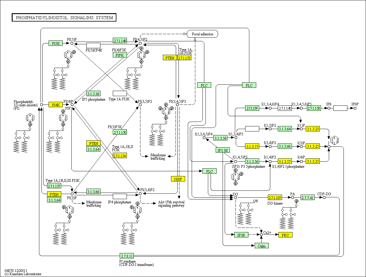
In a recent paper in Cell 143, 897-910 2010, Chakraborty et. al. report a new player in the insulin story.  This is reviewed in the same issue of Cell by Brendan D. Manning Cell 143 861-862 2010 (DOI 10.1016/j.cell.2010.11.040).  They have discovered an insulin receptor product which has not been reported earlier.  Activation of the insulin receptor stimulates both phosphorylation of IRS1 and activation of inositol hexakisphosphate (IP6) kinase (IP6K1).  The latter yields 5-diphospho-inositolpentakisphosphate (5-PP-IP5 or IP7).  IP7 binds to Akt, inactivating it and preventing its function as a substrate for mTORC2* and PDK1 phosphorylation.  That is, insulin produces signal substances that both activate and deactivate Akt.  The "on" signal is the PIP2-PIP3 sequence.  The "off" signal is the IP6-IP7 sequence.  The balance between these may dominate insulin regulation of metabolism.  Perhaps even more important is the possibility that the key to understanding "insulin resistance" may lie in the balance between these. 


*mTOR
mTOR is a nuclear serine/threonine protein kinase found in two complexes (mTORC1 and mTORC2) in most of the body's tissues.  These appear to be essential in the regulation of metabolism, including organization of the insulin and growth factor signals.  mTOR is also an important element in the ageing process and in development of several types of cancer.  There is a very large literature concerning mTOR.  An analysis of this lies beyond the scope of Medbio.info.  However, I acknowledge that mTOR is of major interest for further developments in medicine.  I will advise those who are interested to download two important articles from Wikipedia.  1: http://en.wikipedia.org/wiki/MTOR.  2: http://en.wikipedia.org/wiki/Discovery_and_development_of_mTOR_inhibitors.

PI signalointikartta


Alfa Tokoferoli ja PI voivat vaihtua fosfaatissa

https://globalmedicaldiscovery.com/key-scientific-articles/modulation-phosphorylation-tocopherol-phosphatidylinositol-htap1sec14l2-mediated-lipid-exchange/

Phosphorylated, but not un-phosphorylated tocopherol (vitamin E) induces expression of the vascular endothelial growth factor (VEGF).
 A specific tocopherol and phosphatidylinositol binding protein, hTAP1/SEC14L2, exchanges tocopherol and phosphatidylinositol and modulates their phosphorylation. Lipid binding and exchange by hTAP/SEC14L2 modulates signal transduction and gene expression leading to induction of the expression of VEGF.
A novel signaling pathway involving hTAP1/SEC14L2-mediated lipid exchange lends itself as target for pathogenic and therapeutic studies.
 
Figure Legend
Lipid exchange molecular model for hTAPs and its role in lipid transport and enzyme regulation. (A) hTAPs transfer lipids (e.g. phosphatidylinositol, phosphatidylcholine, vitamin E, squalene) from/to cellular import/export sites or between different membranes and membrane domains such as lipid rafts
. (A and B)
 hTAPs mediated lipid transport may change membrane lipid composition and membrane curvature and in this way influence signal transduction, gene expression and secretion.
 (B) hTAPs bring lipid substrates (S) to specific enzymes (E) (e.g. phosphatidylinositol-3-kinases PI3K , tocopherol kinase, squalene epoxidase), present them in the correct orientation and timing, and/or remove the lipid products (P) from the enzyme, thus enhancing lipid turnover at the catalytic center (CC).
 Lipid release and presentation occurs by lipid exchange with a homotypic or heterotypic lipid and may occur preferentially upon interaction of hTAPs with membranes, thus confining lipid membrane removal/insertion and lipid enzymatic modification to membranes.
 Selected further reading
  •  Kempna P, Zingg JM, Ricciarelli R, Hierl M, Saxena S, Azzi A. 2003. Cloning of novel human SEC14p-like proteins: cellular localization, ligand binding and functional properties. Free Radic. Biol. Med. 34:1458-72
  • Zingg JM, Azzi A, Meydani M. 2014. Induction of VEGF expression by alpha-tocopherol and alpha-tocopheryl phosphate via PI3Kgamma/PKB and hTAP1/SEC14L2-mediated lipid exchange. J Cell Biochem
  • Zingg JM, Libinaki R, Lai CQ, Meydani M, Gianello R, et al. 2010. Modulation of gene expression by alpha-tocopherol and alpha-tocopheryl phosphate in THP-1 monocytes. FRBM 49:1989-2000
  • Zingg JM, Meydani M, Azzi A. 2010. alpha-Tocopheryl phosphate – An active lipid mediator? Mol Nutr Food Res 54:1-14
  • Zingg JM, Meydani M, Azzi A. 2012. alpha-Tocopheryl phosphate-An activated form of vitamin E important for angiogenesis and vasculogenesis? BioFactors 38:24-33

Modulation of phosphorylation of tocopherol and phosphatidylinositol by hTAP1/SEC14L2-mediated lipid exchange.. Global Medical Discovery


Journal Reference

Zingg JM1, Libinaki R2, Meydani M1, Azzi A1. PLoS One. 2014;9(7):e101550.
1Vascular Biology Laboratory, JM USDA-Human Nutr. Res. Ctr. On Aging, Tufts University, Boston, Massachusetts, United States of America.and
2Dept. Biochem. and Mol. Biology, Monash University, Melbourne, VIC, Australia.
Abstract
 The vitamin E derivative, alpha-tocopheryl phosphate ({Alpha}TP), is detectable in cultured cells, plasma and tissues in small amounts, suggesting the existence of enzyme(s) with {Alpha}-tocopherol ({Alpha}T) kinase activity. Here, we characterize the production of {Alpha}TP from {Alpha}T and [{Gamma}-32P]-ATP in primary human coronary artery smooth muscle cells (HCA-SMC) using separation by thin layer chromatography (TLC) and subsequent analysis by Ultra Performance Liquid Chromatography (UPLC). In addition to {Alpha}T, although to a lower amount, also {Gamma}T is phosphorylated. In THP-1 monocytes, {Gamma}TP inhibits cell proliferation and reduces CD36 scavenger receptor expression more potently than {Alpha}TP. Both {Alpha}TP and {Gamma}TP activate the promoter of the human vascular endothelial growth factor (VEGF) gene with similar potency, whereas {Alpha}T and {Gamma}T had no significant effect. The recombinant human tocopherol associated protein 1 (hTAP1, hSEC14L2) binds both {Alpha}T and {Alpha}TP and stimulates phosphorylation of {Alpha}T possibly by facilitating its transport and presentation to a putative {Alpha}T kinase. Recombinant hTAP1 reduces the in vitro activity of the phosphatidylinositol-3-kinase gamma (PI3K{Gamma}) indicating the formation of a stalled/inactive hTAP1/PI3K{Gamma} heterodimer. The addition of {Alpha}T, {Beta}T, {Gamma}T, {delta}T or {Alpha}TP differentially stimulates PI3K{Gamma}, suggesting facilitated egress of sequestered PI from hTAP1 to the enzyme. It is suggested that the continuous competitive exchange of different lipophilic ligands in hTAPs with cell enzymes and membranes may be a way to make these lipophiles more accessible as substrates for enzymes and as components of specific membrane domains.